Acquisition
Parameters
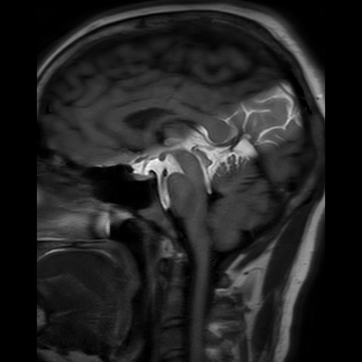
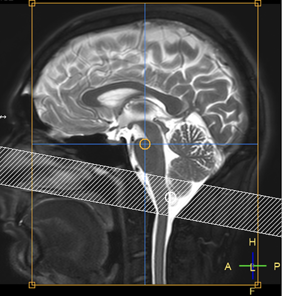
Craniocervical Junction Placement
Download Example Images
The placement of the tag-based imaging plane will be a midline sagittal image with an approximate field of view covering the third ventricle through the C3 cervical vertebrae. The tagging location is placed independently of the imaging plane. The tagging plane should be a slightly oblique axial slice which covers the CSF near the median aperture, without extending into either the central canal or fourth ventricle.
Video of tag at craniocervical junction
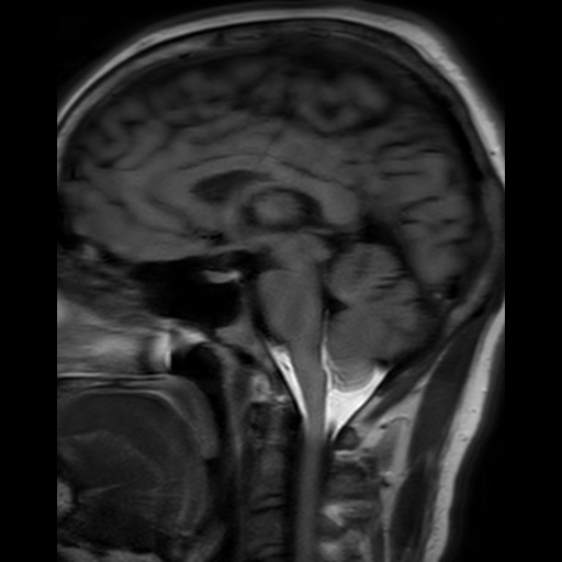
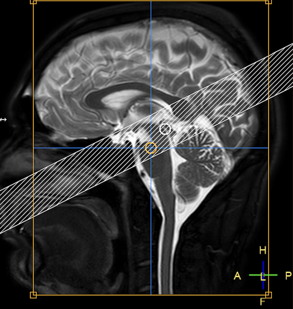
Prepontine Cistern Placement
Download Example Images
The placement of the tag-based imaging plane will be a midline sagittal image with an approximate field of view covering the lateral ventricles through the prepontine cistern. The tagging location is placed independently of the imaging plane. The tagging plane should be a slightly oblique axial slice which covers from the interpeduncular cistern down to approximately the pontine cistern. Note: if the tag extends too far superior to the interpeduncular cistern it might accidentality tag the third ventricle or lateral ventricle, which would make it difficult to interpret if the ETV is patent.
Video of tag at prepontine cistern